


NfL – A Prognostic Biomarker Of Neuroaxonal Damage
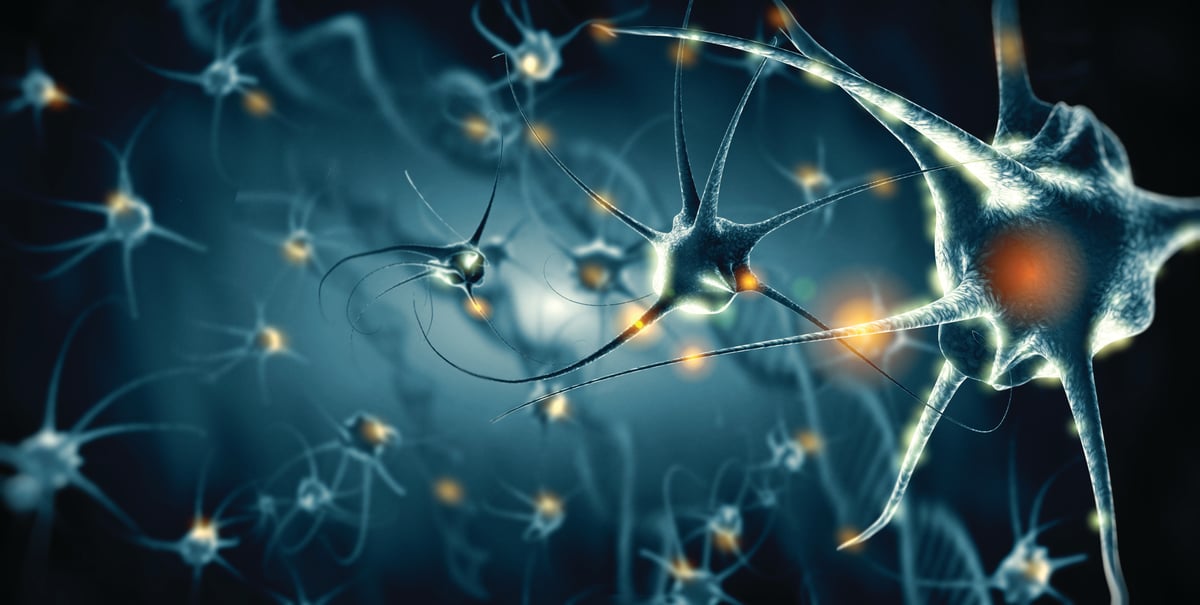
Neurofilaments are parts of the cytoskeletal structure of neurons and are abundantly found in axons. They are important for maintaining size and providing structural support for the neuron. There are currently five known neurofilament proteins that can co-assemble in different combinations, one of which is neurofilament light (NfL).
NfL as a Biomarker of Neuroaxonal Damage
Upon neural damage in the central nervous system (CNS), neurofilament proteins are released into the cerebrospinal fluid (CSF) and can be used as biomarkers for axonal damage and neuronal death. In this context, NfL is the most commonly used biomarker.
There are a number of diseases and disorders where the ability to measure neural damage is essential. NfL has is being used as a marker for disease activity and progression in several neurological diseases such as Alzheimer’s disease, stroke, ALS, frontotemporal dementia and multiple sclerosis. In addition, NfL has been associated with disorders of the paraneoplastic CNS and the peripheral nervous system.
What makes NfL especially interesting is that it is a general marker of neurological damage and is not disease specific. This means that NfL markers can be used in neurological disorders where no good specific markers exist today. Another advantage of using NfL is its flexibility as it can be used as a biomarker for both disease state, prognosis and response to treatment, allowing insight not only in to disease presence but also progression and severity.
From CSF to Blood: Expanding Access to NfL Testing
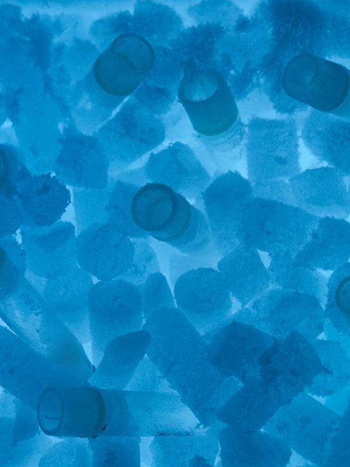
Until recently, most studies on NfL in disease measured the protein in CSF samples. However, CSF sampling is invasive and requires lumbar puncture, which has led scientists to look for a reliable methodology for assessing NfL in other biofluids.
The use of single molecule array (Simoa) - a form of super-sensitive ELISA that can be used to measure very low concentrations of protein - has opened up the possibility of less invasive testing. Measurements of NfL concentration in blood serum using this technique correlate very well with those found in CSF, which allows patient NfL measurements to be taken from simple blood tests.
NfL Testing With Wieslab Diagnostic Services
Wieslab offers clinical testing of NfL across multiple sample types - CSF, serum, and EDTA plasma using Simoa technology. NfL can be tested independently with a regular (7 days) or urgent (48hr) turnaround time, or as part of tailored panels for Inflammatory Neuropathy, ALS, MS, and more.