


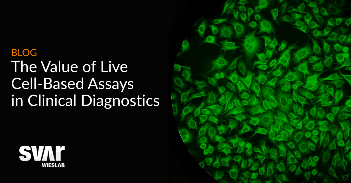

The detection of disease-specific antibodies is at the heart of modern clinical diagnostics for autoimmune and neurological disorders. Over the past decades, cell-based assays (CBAs) have become essential tools for identifying these antibodies using cells expressing target antigen proteins. Today, a growing body of evidence highlights the added value of live cell-based assays (LCBA), a method that more closely reflects biological reality and can improve diagnostic accuracy.
What Is The Current Clinical Standard?
The most widely used format in clinical laboratories today is the fixed cell-based assay (FCBA). FCBA involves transfecting cells to express a specific antigen, fixing them to preserve structure, and then incubating them with a patient sample. If disease-specific antibodies are present, they bind to the antigen and are detected through fluorescence, typically read using manual microscopy.
These assays are commonly used because they are robust, scalable, and compatible with standardized workflows, including compliance with diagnostic regulations such as IVDR.
However, fixation is a harsh chemical process. It can alter the antigen’s conformation on the cell surface, which can be a particular issue for antibodies that only bind when the target proteins are folded in their native state. Scientific studies have shown that this process can significantly reduce the sensitivity of FCBAs, potentially leading to false negatives and missed diagnoses 1.
How Are Live Cell-Based Assays Different?
Live cell-based assays represent an evolution of this technology. While the core steps – transfection, fixation, and sample addition - are the same, the key difference lies in when the patient sample is introduced.
In LCBA, the transfected cells are incubated with the patient sample before any fixation occurs. This means that the antigens stay in their native conformation, allowing antibodies to interact with the cells in an environment that more closely mimics real biological conditions.
Another key difference is in the readout process. Alongside microscopy, LCBAs can also be assessed using flow cytometry which allows quantitative and objective analysis, compared to the more subjective readout provided by microscopy in FCBAs.
.png?width=1200&height=627&name=Live%20CBA%20(1).png)
What Are The Benefits Of LCBA?
The primary advantage of LCBAs lies in their enhanced sensitivity. Multiple clinical studies, particularly in autoimmune neurology, demonstrate that LCBAs can significantly outperform FCBAs on this measure.
In one large real-world study 2, the detection of antibodies against myelin oligodendrocyte glycoprotein (MOG), found sensitivity of approximately 95% with LCBA compared to just 45.7% with FCBA. Similarly, for aquaporin-4 (AQP4) antibodies, sensitivity increased from around 71.6% with FCBA to over 97% when using LCBA.
Importantly, this increased sensitivity does not come at the expense of specificity. Both methods generally demonstrate high specificity, often approaching or reaching 100% for certain targets like AQP4. This means that LCBA can improve detection of true positives without significantly increasing the risk of false positives.
This high sensitivity and specificity is particularly critical in central nervous system (CNS) diseases such as Neuromyelitis optica spectrum disorder (NMOSD) - indicated by AQP4 antibodies - and MOG antibody-associated disease (MOGAD), where early and accurate diagnosis can significantly impact treatment decisions and long-term outcomes.

Wieslab’s Live Cell-Based Assays
Wieslab currently offers LCBA testing for key clinically relevant targets, including Aquaporin-4 (AQP4) antibodies and Myelin oligodendrocyte glycoprotein (MOG) antibodies. These assays are designed to support clinicians in diagnosing complex autoimmune neurological conditions with greater confidence and accuracy. Wieslab’s LCBA platform uses flow cytometry for detection, providing sensitivity, specificity, and objectivity in diagnostics.
Learn more about Wieslab’s live cell-based assays through the links below.